

Isolated visceral arteries dissection: Report of three cases and literature review

| Available Online: | April, 2024 |
| Page: | 55-60 |
Author for correspondence:
Christiana Anastasiadou MD, MSc, PhDc
Papanikolaou Avenue, Chortiatis P.O 57010 Thessaloniki, Greece
E-mail: an.xristiana@hotmail.com
doi: 10.59037/me0nkk18
ISSN 2732-7175 / 2024 Hellenic Society of Vascular and Endovascular Surgery Published by Rotonda Publications All rights reserved. https://www.heljves.com
1 Department of Vascular and Endovascular surgery, “Georgios Papanikolaou” General Hospital of Thessaloniki
2 Radiology Department, “Georgios Papanikolaou” General Hospital of Thessaloniki
3 4th Department of Surgery, Medical School, Aristotle University of Thessaloniki, “Georgios Papanikolaou” General Hospital of Thessaloniki
Abstract
Full Text
References
Images
Abstract
Abstract:
Spontaneous, isolated visceral artery dissection represents an infrequent clinical occurrence. Over the past decade, there has been a notable rise in reported cases within the medical literature. There is no optimal therapy for this condition, nevertheless, conservative therapy and close follow-up for uncomplicated cases is accepted. Computed tomography angiography is the diagnostic tool of choice, whereas blood tests usually have no specifical abnormalities. Nowadays, endovascular therapy is therapy of choice for complicated cases, especially where exploration of the abdomen is not necessary. We report three cases of successful conservative therapy and we present a literature review.
Keywords: visceral, dissection, spontaneous, isolated, celiac artery, superior mesenteric artery
Full Text
Introduction
Isolated visceral artery dissection represents an infrequent clinical occurrence. Over the past decade, there has been a notable rise in reported cases within the medical literature. This increase is primarily ascribed to the growing utilization of computed tomography angiography (CTA), although it may also be linked to an overall elevation in the incidence of visceral artery dissections. The precise pathophysiology of this condition remains unclear. Common risk factors identified among affected individuals encompass smoking and hypertension, implying that atherosclerosis and heightened shear stress may significantly contribute to the pathophysiological mechanisms. In this context, we present three cases of isolated visceral artery dissection managed conservatively, accompanied by a comprehensive literature review elucidating the characteristics of this uncommon medical phenomenon. Informed consent has been obtained from the patients for publication of the case report and accompanying images. Also, approval was obtained from the local ethics committee.
Case 1
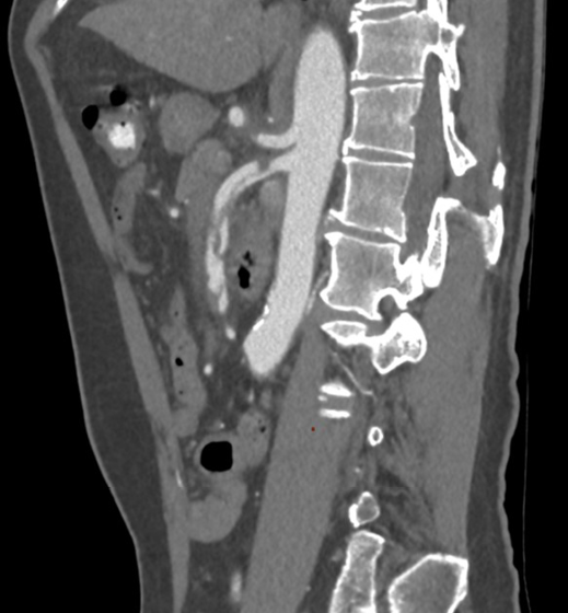
A 52-year old woman presented to the emergency department complaining of abdominal pain radiating to her back. The pain was characterized as postprandial, commencing four weeks prior and intensifying over the last twenty-four hours. Clinical assessment disclosed a supple abdomen with generalized tenderness. The patient’s medical history disclosed inadequately controlled arterial hypertension. Laboratory investigations demonstrated values within the normal range. CTA revealed a celiac artery (CA) dissection with an intimal flap originating approximately 1.2cm from the celiac artery ostium for a length of 1.7cm. (fig.1) Hepatic, splenic and gastric artery were patent with no signs of flow limitation, thrombosis, aneurysm formation or intestinal ischemia. Conservative management was initiated. Subsequently, the pain was effectively alleviated, and the patient has remained asymptomatic for a duration of two years. (fig.2)
Case 2
A-64-year-old man presented to the emergency department with recurrence of his abdominal pain and indigestion. He had been hospitalized two weeks prior at a different medical facility, where he received treatment involving antibiotics and proton pump inhibitors. The patient’s medical background comprised a history of hypertension, coronary artery disease, and diabetes mellitus. Physical examination revealed a diffused, mild abdominal tenderness. Platelets and white blood count deviated from the normal range. X-rays and ultrasonography were normal. CTA revealed stenosis at the celiac artery ostium with post-stenotic dilatation (fig.3) as well as a dissection of superior mesenteric artery (SMA) with a flap originated at 1.2cm from its ostium, with a length of 8.5cm and re-entry point with patent branches of the SMA. (fig.4) The patient was successfully managed conservatively. A CT scan performed three months post the incident exhibited consistent and unchanging results.
Case 3
A 51-year-old man presented to the emergency department reporting acute abdominal and left flank pain. The patient’s medical history encompassed inadequately controlled hypertension, dyslipidaemia, and a history of tobacco use. CTA revealed dissection of the celiac artery and flow limitation to its branches. The patient was admitted to the hospital and was successfully treated conservatively, including bowel rest, antiplatelet and anticoagulation therapy. Ten days later, he underwent a repeated CTA which revealed patent left gastric artery. (fig.5) He remained asymptomatic, with stable CT scan findings even 7 years after this incident.
All three patients were treated conservatively in the acute phase and the therapy included anticoagulation, antiplatelet, b-blocker medications and bowel rest. Enteral nutrition was incrementally introduced starting from the sixth day of their hospitalization . Upon discharge, they were prescribed antiplatelet agents, and the follow-up period ranged from three months to 7 years.
Discussion
Spontaneous dissection of visceral arteries was traditionally considered an exceedingly rare condition, given an estimated incidence of approximately 0.08% whereas synchronous dissection of more than one visceral artery is even more extraordinary.1-7 A comprehensive review of published articles within the PubMed database (2012-2022) was conducted to investigate isolated visceral artery dissection. The search yielded information on more than 600 cases. The augmented identification of spontaneous visceral artery dissection is attributed either to a genuine increase in the prevalence of such cases or to the enhanced detectability, facilitated by contemporary imaging modalities.8 The demographic profile of affected individuals typically comprises middle-aged, Asian men. Upon presentation, these patients commonly exhibit a history of smoking and hypertensive urgency.2,4,9-12 The superior mesenteric artery is most often affected, with the celiac artery following as the next most commonly affected vessel.10,13,14
Bax et al, in a very interesting article, discuss on pathophysiology of arterial dissections.15 Various factors including hypertension, age, gender, connective tissue diseases, atherosclerosis, arterial cystic necrosis, trauma and smoking are considered to be associated with the disease. An intriguing pathophysiologic mechanism was suggested by Wu et al, proposing that SMA is more susceptible to shear stress in the transition zone from fixed retropancreatic to relatively mobile segment in the mesenteric root, analogous to what is seen at the ligamentum arteriosum in thoracic aortic dissection.16 Celiac artery dissection should include investigation for median arcuate ligament syndrome.17-19 An additional finding identified in this study, is that pancreatic enzymes released during pancreatitis can erode adjacent arterial wall and thus, the presence of acute pancreatitis might have triggered arterial dissection.6,20
Clinical presentation most often includes acute abdominal or flank pain. However, it could be present in a more indolent fashion, with an insidious onset, lasting days or weeks before the pain becomes more severe.5 Yamaguchi et al reported a case where a patient presented with acalculous cholecystitis as a result of hepatomesenteric trunk dissection.21 The pain can be postprandially or irrelevant with food intake, and the character may be sharp or dull. Some patients remained totally asymptomatic, and they were diagnosed incidentally or they were misdiagnosed.5,14,19,22 Abdominal pain may be implying bowel ischemia, perforation and peritonitis or aneurysmal formation with imminent rupture of the artery. Nevertheless, the abdominal pain could alternatively be explained as an inflammatory response triggered by the dissection, thereby provoking pain through stimulation of the visceral nerve plexus. Current literature supports that the degree of pain is positively correlated with the length of the dissected blood vessel.23 An author suggests that there were no significant differences in medical history or medications between symptomatic and asymptomatic patients. However, patients with abdominal symptoms tended to be younger and were more frequently hospitalized.24 Nevertheless, in another study, it was noted that asymptomatic patients were younger (53.9±11.4 vs 58.7±11.2, p = 0.032) and that no significant differences were presented between the artery which was involved (CA vs SMA) in patients with or without symptoms, however there was a trend towards SMA involvement in symptomatic patients (23 (46%) vs 7 (26%), p = 0.085).25
The literature documents that many patients were misdiagnosed. Radiologic modalities are determinant of an accurate diagnosis.26 CT angiography is the preferred diagnostic tool, as it is a rapid, non-invasive, and high resolution examination, which contributes to the visualization of the vessels and of complications from abdominal organs such as necrosis and perforation.5,13 Regarding laboratory blood tests, coagulation markers, such as fibrin degradation products, are known to increase in acute aortic dissection. However, these markers were not markedly elevated in this condition, even in symptomatic patients, possibly because the amount of thrombus in SIVAD is smaller, due to the size of the vessel.24 According to the same study, there were significant differences between symptomatic and asymptomatic patients in white blood cell count and creatine kinase levels, but not in FDPs or d-dimer levels.24
There are no specific guidelines regarding the ideal treatment of visceral dissection. However, a reasonable algorithm is the following: Surgery (open/endo/hybrid) is recommended if the patient in the acute phase presents with rupture, signs of end-organ ischemia, enlargement of the artery (>2cm) or blood flow limitation, in correlation with pain not responding to medication. If none of these conditions exists, the physician can choose conservative therapy (medication, bowel rest). Our strategy includes bowel rest for 5 to 6 days, staged nutrition, anti-hypertensive medication, antiplatelet therapy, low molecular weight heparin at a prophylactic dose, repeated laboratory blood tests including arterial blood gases, and CT angiography on the day of the admission, before discharge, at 3 and at 12 months after patients’ discharge. After the first year, patients are followed up with duplex scan in turn with CT scan to avoid exposure to radiation. Although there is an ongoing debate regarding the efficacy of antithrombotic therapy (anticoagulation and antiplatelet medication), studies revealed that they do not demonstrate any advantages in terms of clinical or morphological outcomes.7,27 Endovascular therapy includes bare metal stent, coil assisting bare stent therapy, coil embolization3,6,18,28,29, whereas open surgical therapy is preferred in cases where exploration of the abdomen is mandatory. Patch angioplasty or bypass is still an option, but endovascular therapy is the preferred method because of its high technical success and low complication rate.30 Nowadays, hybrid approach is almost always available, however there is still no case report announced in Pubmed database.
Regarding prognosis, dissection can progress in various ways. It may exhibit a self-limited course with symptom resolution, progress to involve distal branches, develop into aneurysmal dilatation, or, in more severe cases, culminate in rupture.19 Current literature supports that approximately 20% of patients who were treated conservatively, developed aneurysmal dilatation requiring intervention during the follow-up period.31,32 In a review by Wang it is reported that 8% of the symptomatic celiac artery dissection patients and 12% of the symptomatic superior mesenteric artery dissection patients who were managed conservatively, required secondary intervention during follow-up, whereas none of the asymptomatic patients needed further intervention.33 Superior mesenteric artery seems to fail to achieve complete remodeling, and therefore, it is correlated with more complications.33 Moreover, comparing visceral artery dissection with renal artery dissection it can be supported that the latter has worst prognosis, since it is correlated with increased complications and mortality.34,35 Patients who have visceral artery dissection with otherwise normal appearing arteries carry a higher risk of major adverse arterial events compared with those with fibromuscular dysplasia, primarily because of recurrent dissections.36,37
In conclusion, given the heightened frequency of case series and reports in the literature over the past decade, one may infer a probable contemporary escalation in the prevalence of isolated visceral artery dissection. This condition poses challenges in the emergency department where its symptoms, encompassing abdominal and back pain, are commonplace, and clinical presentation may mimic other gastrointestinal or musculoskeletal disorders, potentially leading to misdiagnosis. While conservative management during the acute phase is often feasible, further comprehensive data on mid and long-term outcomes and management are needed.
Acknowledgments
None.
Conflicts of interest
The authors declare no conflicts of interest.
References
- Le TB, Jeon YS, Hong KC, Cho SG, Park KM. Spontaneous dissections of multiple visceral arteries: An extremely rare case. Ann Surg Treat Res. 2017;92(4):225-229. doi:10.4174/astr.2017.92.4.225
- Pateman Aciu S, Petrochko J, Bassik N, Fisher J. Spontaneous isolated celiac and splenic artery dissection with splenic infarction. Radiol Case Rep. 2022;17(6):2085-2091. doi:10.1016/j.radcr.2022.03.060
- Moran J, Galla N, Ranade M. Endovascular Stenting in a Rare Case of Multiple Spontaneous Visceral Arterial Dissections. Vasc Endovascular Surg. 2021;55(3):269-272. doi:10.1177/1538574420954574
- Amitai Komem D, Sukenik Halevy R, Griton Y, et al. A Rare Case of 7 Simultaneous Arterial Dissections and Review of The Literature. Vasc Endovascular Surg. 2019;53(7):617-622. doi:10.1177/1538574419864783
- Lalani K, Devasia T, Paramasivam G. Spontaneous dissection of coeliac and superior mesenteric artery: Double whammy. BMJ Case Rep. 2021;14(3). doi:10.1136/bcr-2020-240047
- Yamaguchi H, Murata S, Onozawa S, Sugihara F, Hayashi H, Kumita S ichiro. Strategy for the treatment of spontaneous isolated visceral artery dissection. Eur J Radiol Open. 2019;6:9-15. doi:10.1016/j.ejro.2018.11.003
- Heo SH, Kim YW, Woo SY, Park YJ, Park KB, Kim DK. Treatment strategy based on the natural course for patients with spontaneous isolated superior mesenteric artery dissection. In: Journal of Vascular Surgery. Vol 65. Mosby Inc.; 2017:1142-1151. doi:10.1016/j.jvs.2016.10.109
- Gao F, Huang X, Ren D, Wang Y, Guo J, Deng G. Results Obtained with the Protege EverFlex Self-expanding Bare Stent in Interventional Treatment of Spontaneous Isolated Visceral Artery Dissection. Ann Vasc Surg. 2021;77:86-93. doi:10.1016/j.avsg.2021.05.017
- Ichiba T, Naito H, Nagata T, Masuda R, Hata M, Maeda K. Spontaneous isolated left gastric artery dissection: unusual visceral artery dissection. Acute Medicine & Surgery. 2016;3(4):369-371. doi:10.1002/ams2.191
- Shiraki H, Kasamoto M, Yasutomi M, et al. Clinical Features of Spontaneous Isolated Dissection of Abdominal Visceral Arteries. J Clin Med Res. 2020;12(1):13-17. doi:10.14740/jocmr3916
- Alcantara S, Yang CK, Sasson J, et al. The evidence for nonoperative management of visceral artery dissections: A single-center experience. In: Annals of Vascular Surgery. Vol 29. Elsevier Inc.; 2015:103-108. doi:10.1016/j.avsg.2014.09.004
- Cavalcante RN, Motta-Leal-Filho JM, De Fina B, et al. Systematic literature review on evaluation and management of isolated spontaneous celiac trunk dissection. Ann Vasc Surg. 2016;34:274-279. doi:10.1016/j.avsg.2015.12.009
- He Q, Yu F, Fu Y, et al. Evaluation of isolated abdominal visceral artery dissection with multi-scale spiral computed tomography: a retrospective case series. J Cardiothorac Surg. 2021;16(1). doi:10.1186/s13019-021-01428-8
- Nonami S, Nakanishi T, Tanizaki S, et al. Characteristics and diagnostic pitfalls of spontaneous visceral artery dissection in the emergency department. American Journal of Emergency Medicine. 2016;34(6):1092-1096. doi:10.1016/j.ajem.2016.02.073
- Bax M, Romanov V, Junday K, et al. Arterial dissections: Common features and new perspectives. Front Cardiovasc Med. 2022;9. doi:10.3389/fcvm.2022.1055862
- Wu Z, Yi J, Xu H, et al. The Significance of the Angle between Superior Mesenteric Artery and Aorta in Spontaneous Isolated Superior Mesenteric Artery Dissection. Ann Vasc Surg. 2017;45:117-126. doi:10.1016/j.avsg.2017.06.156
- Takayama T, Miyata T, Shirakawa M, Nagawa H. Isolated spontaneous dissection of the splanchnic arteries. J Vasc Surg. 2008;48(2):329-333. doi:10.1016/j.jvs.2008.03.002
- Yamaguchi H, Murata S, Ueda T, et al. New technique for false lumen coiling of spontaneous isolated superior mesenteric artery dissection. CVIR Endovasc. 2021;4(1). doi:10.1186/s42155-021-00225-7
- Otsuka H, Sato T, Aoki H, Nakagawa Y, Inokuchi S. Optimal management strategy for spontaneous isolated dissection of a visceral artery. Vascular. 2018;26(2):169-174. doi:10.1177/1708538117722879
- Black TP, Obando J V., Burbridge RA. Pancreatitis Secondary to Celiac Trunk Dissection. ACG Case Rep J. 2014;1(2):106-108. doi:10.14309/crj.2014.16
- Yamaguchi S, Kitazono K, Kamiyama T, Ohishi M. Hepatomesenteric Trunk Dissection Complicated with Acalculous Cholecystitis. Internal Medicine. 2023;62(15):2293-2294. doi:10.2169/internalmedicine.1004-22
- Ko SH, Hye R, Frankel DA. Management of spontaneous isolated visceral artery dissection. Ann Vasc Surg. 2015;29(3):470-474. doi:10.1016/j.avsg.2014.10.026
- Yun WS, Kim YW, Park KB, et al. Clinical and Angiographic Follow-up of Spontaneous Isolated Superior Mesenteric Artery Dissection. European Journal of Vascular and Endovascular Surgery. 2009;37(5):572-577. doi:10.1016/j.ejvs.2008.12.010
- Tanaka Y, Yoshimuta T, Kimura K, et al. Clinical characteristics of spontaneous isolated visceral artery dissection. J Vasc Surg. 2018;67(4):1127-1133. doi:10.1016/j.jvs.2017.08.054
- Morgan CE, Mansukhani NA, Eskandari MK, Rodriguez HE. Ten-year review of isolated spontaneous mesenteric arterial dissections. J Vasc Surg. 2018;67(4):1134-1142. doi:10.1016/j.jvs.2017.08.071
- Hoglund JR, Blackwell JH, Gibbs MA. Spontaneous celiac artery dissection. American Journal of Emergency Medicine. 2020;38(7):1545.e3-1545.e5. doi:10.1016/j.ajem.2020.04.041
- Melnychuk E, Strony R. Spontaneous Isolated Visceral Artery Dissection in a Middle Aged Male. Case Rep Emerg Med. 2017;2017:1-3. doi:10.1155/2017/3704348
- Perini P, Baque J, Chau Y, Sedat J, Batt M. Percutaneous embolization of symptomatic dissecting aneurysms of the celiac artery. Acta radiol. 2014;55(9):1076-1081. doi:10.1177/0284185113511079
- Mkangala AM, Liang H, Dong XJ, Su Y, HaoHao L. Safety and efficacy of conservative, endovascular bare stent and endovascular coil assisting bare stent treatments for patients diagnosed with spontaneous isolated superior mesenteric artery dissection. Wideochirurgia I Inne Techniki Maloinwazyjne. 2020;15(4):608-619. doi:10.5114/WIITM.2020.92403
- Zhu Y, Peng Y, Xu M, et al. Treatment Strategies and Outcomes of Symptomatic Spontaneous Isolated Superior Mesenteric Artery Dissection: A Systematic Review and Meta-analysis. Journal of Endovascular Therapy. 2018;25(5):640-648. doi:10.1177/1526602818796537
- Sun J, Li D lin, Wu Z heng, He Y yan, Zhu Q qian, Zhang H kun. Morphologic findings and management strategy of spontaneous isolated dissection of the celiac artery. J Vasc Surg. 2016;64(2):389-394. doi:10.1016/j.jvs.2015.12.050
- Galastri FL, Cavalcante RN, Motta-Leal-Filho JM, et al. Evaluation and management of symptomatic isolated spontaneous celiac trunk dissection. In: Vascular Medicine (United Kingdom). Vol 20. SAGE Publications Ltd; 2015:358-363. doi:10.1177/1358863X15581447
- Wang J, He Y, Zhao J, et al. Systematic review and meta-analysis of current evidence in spontaneous isolated celiac and superior mesenteric artery dissection. J Vasc Surg. 2018;68(4):1228-1240.e9. doi:10.1016/j.jvs.2018.05.014
- Yokoyama Y, Nakajima M. Spontaneous Renal Artery Dissection in a Man with Previous Spontaneous Superior Mesenteric Artery Dissection. Case Rep Vasc Med. 2020;2020:1-4. doi:10.1155/2020/4726381
- Jeong MJ, Kwon H, Kim A, et al. Clinical Outcomes of Conservative Treatment in Patients with Symptomatic Isolated Spontaneous Renal Artery Dissection and Comparison with Superior Mesenteric Artery Dissection. European Journal of Vascular and Endovascular Surgery. 2018;56(2):291-297. doi:10.1016/j.ejvs.2018.05.002
- Henkin S, Wysokinski WE, Tweet M, et al. Spontaneous visceral artery dissections in otherwise normal arteries: Clinical features, management, and outcomes compared with fibromuscular dysplasia. In: Journal of Vascular Surgery. Vol 73. Mosby Inc.; 2021:516-523.e2. doi:10.1016/j.jvs.2020.05.068
- Su KYC, Stanhope ML, Kaufman BPW. Spontaneous hepatic artery dissection-A rare presentation of fibromuscular dysplasia. Oxf Med Case Reports. 2016;2016(11):273-278. doi:10.1093/omcr/omw083


